Vacuole size control: regulation of PtdIns(3,5)P2 levels by the vacuole-associated Vac14-Fig4 complex, a PtdIns(3,5)P2-specific phosphatase
- PMID: 14528018
- PMCID: PMC307524
- DOI: 10.1091/mbc.e03-05-0297
Vacuole size control: regulation of PtdIns(3,5)P2 levels by the vacuole-associated Vac14-Fig4 complex, a PtdIns(3,5)P2-specific phosphatase
Abstract
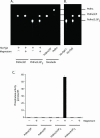
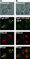
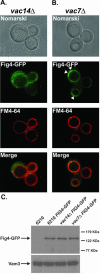
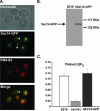
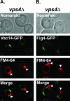
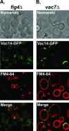
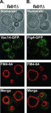
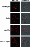
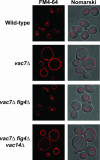
In the budding yeast Saccharomyces cerevisiae, phosphatidylinositol 3,5-bisphosphate (PtdIns(3,5)P2) is synthesized by a single phosphatidylinositol 3-phosphate 5-kinase, Fab1. Cells deficient in PtdIns(3,5)P2 synthesis exhibit a grossly enlarged vacuole morphology, whereas increased levels of PtdIns(3,5)P2 provokes the formation of multiple small vacuoles, suggesting a specific role for PtdIns(3,5)P2 in vacuole size control. Genetic studies have indicated that Fab1 kinase is positively regulated by Vac7 and Vac14; deletion of either gene results in ablation of PtdIns(3,5)P2 synthesis and the formation of a grossly enlarged vacuole. More recently, a suppressor of vac7Delta mutants was identified and shown to encode a putative phosphoinositide phosphatase, Fig4. We demonstrate that Fig4 is a magnesium-activated PtdIns(3,5)P2-selective phosphoinositide phosphatase in vitro. Analysis of a Fig4-GFP fusion protein revealed that the Fig4 phosphatase is localized to the limiting membrane of the vacuole. Surprisingly, in the absence of Vac14, Fig4-GFP no longer localizes to the vacuole. However, Fig4-GFP remains localized to the grossly enlarged vacuoles of vac7 deletion mutants. Consistent with these observations, we found that Fig4 physically associates with Vac14 in a common membrane-associated complex. Our studies indicate that Vac14 both positively regulates Fab1 kinase activity and directs the localization/activation of the Fig4 PtdIns(3,5)P2 phosphatase.
Figures


Similar articles
-
Assembly of a Fab1 phosphoinositide kinase signaling complex requires the Fig4 phosphoinositide phosphatase.Mol Biol Cell. 2008 Oct;19(10):4273-86. doi: 10.1091/mbc.e08-04-0405. Epub 2008 Jul 23. Mol Biol Cell. 2008. PMID: 18653468 Free PMC article.
-
Regulation of Fab1 phosphatidylinositol 3-phosphate 5-kinase pathway by Vac7 protein and Fig4, a polyphosphoinositide phosphatase family member.Mol Biol Cell. 2002 Apr;13(4):1238-51. doi: 10.1091/mbc.01-10-0498. Mol Biol Cell. 2002. PMID: 11950935 Free PMC article.
-
Vac14 controls PtdIns(3,5)P(2) synthesis and Fab1-dependent protein trafficking to the multivesicular body.Curr Biol. 2002 Jun 4;12(11):885-93. doi: 10.1016/s0960-9822(02)00891-6. Curr Biol. 2002. PMID: 12062051
-
Yeast vacuole inheritance and dynamics.Annu Rev Genet. 2003;37:435-60. doi: 10.1146/annurev.genet.37.050203.103207. Annu Rev Genet. 2003. PMID: 14616069 Review.
-
Phosphatidylinositol-3,5-bisphosphate: no longer the poor PIP2.Traffic. 2012 Jan;13(1):1-8. doi: 10.1111/j.1600-0854.2011.01246.x. Epub 2011 Jul 27. Traffic. 2012. PMID: 21736686 Review.
Cited by
-
In vivo, Pikfyve generates PI(3,5)P2, which serves as both a signaling lipid and the major precursor for PI5P.Proc Natl Acad Sci U S A. 2012 Oct 23;109(43):17472-7. doi: 10.1073/pnas.1203106109. Epub 2012 Oct 9. Proc Natl Acad Sci U S A. 2012. PMID: 23047693 Free PMC article.
-
Synthesis and function of membrane phosphoinositides in budding yeast, Saccharomyces cerevisiae.Biochim Biophys Acta. 2007 Mar;1771(3):353-404. doi: 10.1016/j.bbalip.2007.01.015. Epub 2007 Feb 6. Biochim Biophys Acta. 2007. PMID: 17382260 Free PMC article. Review.
-
Endosomal phosphoinositides and human diseases.Traffic. 2008 Aug;9(8):1240-9. doi: 10.1111/j.1600-0854.2008.00754.x. Epub 2008 Apr 21. Traffic. 2008. PMID: 18429927 Free PMC article. Review.
-
Pathogenic mechanism of the FIG4 mutation responsible for Charcot-Marie-Tooth disease CMT4J.PLoS Genet. 2011 Jun;7(6):e1002104. doi: 10.1371/journal.pgen.1002104. Epub 2011 Jun 2. PLoS Genet. 2011. PMID: 21655088 Free PMC article.
-
FIG4 regulates lysosome membrane homeostasis independent of phosphatase function.Hum Mol Genet. 2016 Feb 15;25(4):681-92. doi: 10.1093/hmg/ddv505. Epub 2015 Dec 11. Hum Mol Genet. 2016. PMID: 26662798 Free PMC article.
References
-
- Babst, M., Katzmann, D.J., and Emr, S.D. (2002). ESCRT III: an endosomal-associated heteroologomeric protein complex required for MVB sorting. Dev. Cell 3, 271–282. - PubMed
-
- Beeler, T., Bruce, K., and Dunn, T. (1997). Regulation of cellular Mg2+ by Saccharomyces cerevisiae. Biochim. Biophys. Acta 1323, 310–318. - PubMed
Publication types
MeSH terms
Substances
Grants and funding
LinkOut - more resources
Full Text Sources
Other Literature Sources
Molecular Biology Databases

