. 2023 Oct;55(10):1632-1639.
doi: 10.1038/s41588-023-01505-9. Epub 2023 Sep 18.

Molecular basis for maternal inheritance of human mitochondrial DNA

Affiliations

Molecular basis for maternal inheritance of human mitochondrial DNA

William Lee et al. Nat Genet. 2023 Oct.

Abstract

Uniparental inheritance of mitochondrial DNA (mtDNA) is an evolutionary trait found in nearly all eukaryotes. In many species, including humans, the sperm mitochondria are introduced to the oocyte during fertilization1,2. The mechanisms hypothesized to prevent paternal mtDNA transmission include ubiquitination of the sperm mitochondria and mitophagy3,4. However, the causative mechanisms of paternal mtDNA elimination have not been defined5,6. We found that mitochondria in human spermatozoa are devoid of intact mtDNA and lack mitochondrial transcription factor A (TFAM)-the major nucleoid protein required to protect, maintain and transcribe mtDNA. During spermatogenesis, sperm cells express an isoform of TFAM, which retains the mitochondrial presequence, ordinarily removed upon mitochondrial import. Phosphorylation of this presequence prevents mitochondrial import and directs TFAM to the spermatozoon nucleus. TFAM relocalization from the mitochondria of spermatogonia to the spermatozoa nucleus directly correlates with the elimination of mtDNA, thereby explaining maternal inheritance in this species.


Conflict of interest statement

Competing interests:

Authors declare that they have no competing interests.

Figures

Extended Data Fig. 1.
Extended Data Fig. 1.. Mitochondria of human spermatozoa contain no mtDNA.
A. Schematics of the mtDNA copy number assessment in sperm cells using Whole Genome Sequencing (WGS). B. Box plots of mtDNA copy number (mtDNA/per cell) analysis in bulk spermatozoa and blood samples (n=3); n represents a biologically independent number of samples. Center line, median; box bounds, 25th and 75th percentiles; whiskers, minimum to maximum within 1.5 interquartile range; data points outside whiskers, outliers. C. Illustration of a mitochondrial genome coverage breadth in bulk blood cells (top) and spermatozoa (bottom) sample (donor 2) enriched for mtDNA using long-range PCR amplifying full-length mtDNA in one reaction. A peak in the read density observed in the region of priming of the PCR primers (black arrows, position ~2,000 nt) in spermatozoa but not in the blood cells likely reflects the presence of degraded mtDNA molecules. D. Schematic of human mtDNA showing the location of sequences targeted by different primer pairs. Pink arrows indicate the mtDNA common deletion region. The name and location of the amplicons are shown in blue. E. Representative one-dimension droplet scatter plot illustrating the accuracy of ddPCR in mtDNA detection. The pJET plasmid containing only the mt64-ND1 target mtDNA fragment was used; amplification was performed using mt64-ND1 and mt79-COX3 primers. F. Representative one-dimension droplet scatter plots were obtained using different primer pairs after ddPCR of DNA in the same spermatozoa sample. Blue dots above the green line indicate amplicon-positive droplets. The absolute mtDNA copy number is given for each primer pair at the top of the panel.
Extended Data Fig. 2.
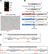
Extended Data Fig. 2.. Mapping of the UTRs of the sperm TFAM isoform.
A. Western blot analysis of the proteins indicated in mitochondria of HEK cells and spermatozoa (SPZ). B. Schematics of the RACE experiment. C. The sequence of the 3’ UTR of the sperm TFAM cDNA D. Schematic illustration of polyadenylation of the TFAM mRNA in somatic cells. PAS – polyadenylation signal. E. Schematic illustration of polyadenylation of the TFAM mRNA in sperm cells. Putative polyadenylation signal (PAS) and UGUAN elements are indicated.
Extended Data Fig. 3.
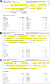
Extended Data Fig. 3.. Identification of TFAM peptides by LC-MS/MS analysis in HEK cells (A), human (B), and Rhesus monkey (C) spermatozoa.
Peptide search was done using MaxQuant. Note that the SGAELCSGCGSR peptide was identified in the human sperm TFAM sample using pFind and, therefore, not shown in panel B.
Extended Data Fig. 4.
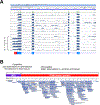
Extended Data Fig. 4.. Pattern of TFAM expression in different human tissues.
A. Exon IT is detected only in mRNA ftom testis. Histograms of RNA-seq data from 10 different tissues have been visualized using the Genome Data Viewer (NCBI). The schematic of the TFAM pre-mRNA is shown below the histograms. B. TFAM peptides matching the mitochondrial pre-sequence are not detected in somatic tissues. The uniquely-mapping protein-calling peptides are shown as blue bars and multi-mapping and non-tryptic peptides are yellow bars. Light blue, 1–4 observations. Dark blue, five or more observations. Proteomic data were obtained from 1099 proteomic experiments. Source: Peptide Atlas (db.systemsbiology.net).
Extended Data Fig. 5.
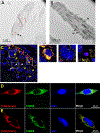
Extended Data Fig. 5.. TFAM localization in spermatozoa and somatic cells.
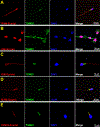
A. Cryo immunogold electron microscopy of spermatozoa head. Red arrows indicate gold particles. A - acrosome. B. Cryo immunogold electron microscopy of spermatozoon midpiece. M- mitochondria. C. Staining of human testicular tissue. Merge image. Red- staining with anti-TFAM antibody, blue- DAPI, green -staining with anti-TOM20 antibody. Red arrows - spermatogonia, yellow – spermatocytes, white – spermatids. The basement membrane is indicated by a dashed line, L- seminiferous tubule lumen. Close-up images of spermatogonia (1), spermatocytes (2) and spermatids (3) correspond to the white squares indicated. D. Over-expressed TFAM having the somatic 5’ and 3’ UTRs shows mitochondrial localization in HeLa cells. E. Overexpressed TFAM lacking UTRs shows mitochondrial localization in HeLa cells
Extended Data Fig. 6.
Extended Data Fig. 6.. Expression of TFAM, H2B, TEFM, and TOM20 in somatic cells and mature spermatozoa.
A. Over-expression of TFAM mRNA having sperm 3’ and 5’ UTR regions result in cytoplasmic localization of this protein in spermatozoa. B. Over-expression of H2B in HeLa cells shows nuclear localization. C. Over-expression of H2B in sperm cells results in cytoplasmic localization. D. Over-expression of TEFM shows mitochondrial localization in spermatozoa. E. Over-expression of TOM20 results in mitochondrial localization in spermatozoa
Extended Data Fig. 7.
Extended Data Fig. 7.. Trafficking of the TFAM variants in HeLa cells.
A. MTSpol-mScarlet protein is localized to the sperm mitochondria. B. MTSpol-mScarlet is localized to mitochondria in HeLa cells. C. MTSpol-Δ42TFAM is localized to the nucleus of HeLa cells. D. MTSTFAM-Scarlet is localized to mitochondria in HeLa cells
Extended Data Fig. 8.
Extended Data Fig. 8.. Expression of the phosphomimicking TFAM variant in HEK293 cells.
A. TFAMS31AA/S34AA-mScarlet protein is localized to the mitochondria of HEK293 cells. B. TFAMS31DD/S34DD-mScarlet protein is localized to the nucleus of HEK293 cells.
Figure 1.
Figure 1.. Mitochondria of human spermatozoa contain no mtDNA.
A. Schematics of the mtDNA copy number measurements in sperm cells by ddPCR. B, C. Representative 1D droplet scatter plots of ddPCR analysis of spermatozoa samples. The multiplex analysis of amplification of two single-copy nuclear genes, TATA-box binding protein 1 and TEFM (B), and two mitochondrial genes, cytochrome b and ND1 (C), in the same spermatozoa sample, is presented. Blue dots above the thresholds (dotted green lines) indicate droplets positive for the target amplicons. D. Summary data indicating mtDNA copy number per cell. E. In situ RNAScope labeling of mtDNA in developing sperm cells in seminiferous tubules of testicular tissue. MtDNA- brown, white arrows - spermatogonia, yellow – spermatocytes, black – spermatozoa. L – seminiferous tubule lumen.
Figure 2.
Figure 2.. Mature sperm cells contain a specific isoform of TFAM.
A. Western blot of mitochondrial lysate of HEK293 cells (lane 1), spermatozoa (lane 2), and spermatozoa in which the tails/midpieces have been removed (lane 3) are shown. Bottom - Western blot of the same gel stained with the anti-VDAC antibody. B. Sperm isoform of TFAM is a product of alternative splicing of the TFAM pre-mRNA. Northern blot of mRNA extracted from sperm (SPZ) or HEK293 cells. C. Schematic illustration of TFAM mRNA structure in somatic and sperm cells. D. Sperm TFAM isoform contains a mitochondrial pre-sequence, as revealed by LC-MS/MS data. MTS -mitochondrial targeting sequence. Tryptic peptides detected in spermatozoa samples are shown below the N-terminal sequence of TFAM.
Figure 3.
Figure 3.. Mature sperm cells contain TFAM in the nucleus but not in the mitochondria.
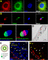
A, B. Confocal microscopy reveals the localization of the endogenous TFAM (red) in mitochondria of HeLa cells (A) and the head of spermatozoa (B). Mitochondria staining with an anti-TOM20 antibody (green) and nucleus - DAPI (blue). C. Deconvolved 3D reconstructed confocal Z-stack image of spermatozoa. Staining with the anti-acrosin antibody (left) or anti-acrosin and anti-TFAM antibody (right). D. Cryo immunogold electron microscopy to reveal localization of TFAM in spermatozoa. The head region is shown. Red arrows indicate gold particles. A - acrosome, NC- nucleus. E. Schematic drawing of a transversal cut of a seminiferous tubule. F, G. Human testicular tissue staining using DAPI (blue, F) and an anti-TFAM antibody (red, G). Red arrows - spermatogonia, yellow – spermatocytes, white – spermatids. The basement membrane is indicated by a dashed line. L – seminiferous tubule lumen.
Figure 4.
Figure 4.. MTS, but not UTR of TFAM mRNA, plays a role in TFAM localization.
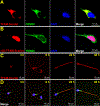
A. Confocal microscopy of HeLa cells transduced with the TFAM-mScarlet fusion having the nuclear 3’ and 5’ UTRs. B. Confocal microscopy of HeLa cells transduced with TFAM-mScarlet fusion lacking the first 42 amino acids. C, D. Confocal microscopy of spermatozoa transduced with TFAM-mScalter protein. Mitochondria staining with an anti-TOM20 antibody (green), nucleus - DAPI (blue), TFAM-mScarlet – red.
Figure 5.
Figure 5.. TFAM import to mitochondria is prevented in spermatozoa.
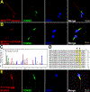
A. Confocal microscopy of spermatozoa transduced with MTSTFAM-mScarlet protein B. Confocal microscopy of spermatozoa transduced with MTSpol-Δ42TFAM-mScarlet C. S34 residue in the spermatozoa TFAM pre-sequence is phosphorylated. The MS/MS spectrum confirming the presence of Ser in TFAM is shown. m/z, mass-to-charge ratio. D. Conservation of the serine residues implicated in TFAM phosphorylation in mammalian species. E. Confocal microscopy of spermatozoa transduced with TFAMS31A/S34A-mScarlet protein
Figure 6.
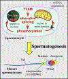
Figure 6.. Sperm TFAM re-localization during spermatogenesis.
During spermatogenesis, TFAM is phosphorylated and prevented from being imported to mitochondria, resulting in mtDNA degradation. Instead, TFAM is accumulated in the nucleus of mature spermatozoa.

Similar articles

Cited by

References

    1. Ankel-Simons F & Cummins JM Misconceptions about mitochondria and mammalian fertilization: implications for theories on human evolution. Proc Natl Acad Sci U S A 93, 13859–63 (1996). - PMC - PubMed
    1. Wallace DC Why do we still have a maternally inherited mitochondrial DNA? Insights from evolutionary medicine. Annu Rev Biochem 76, 781–821 (2007). - PubMed
    1. Sutovsky P et al. Ubiquitinated sperm mitochondria, selective proteolysis, and the regulation of mitochondrial inheritance in mammalian embryos. Biol Reprod 63, 582–90 (2000). - PubMed
    1. Sato M & Sato K Degradation of paternal mitochondria by fertilization-triggered autophagy in C. elegans embryos. Science 334, 1141–4 (2011). - PubMed
    1. Luo SM et al. Unique insights into maternal mitochondrial inheritance in mice. Proc Natl Acad Sci U S A 110, 13038–43 (2013). - PMC - PubMed

Publication types